Revolutionizing Histopathology Lab Workflows: A Digital Transformation StoryLast Tuesday morning started like any other in our busy histopathology lab—until it didn't. At 9:15 AM, we received an urgent biopsy from the OR that needed immediate analysis. In the old days, this would have meant a frantic search for the right glass slide, hoping it wasn't damaged, calling in additional staff, and potentially delaying the surgical procedure. Instead, within 12 minutes, I was reviewing high-resolution digital images from my office while simultaneously consulting with an oncology specialist in Chennai. The surgeon received the critical diagnosis without leaving the OR.
This transformation didn't happen overnight, but it's completely changed how we operate our histopathology lab.
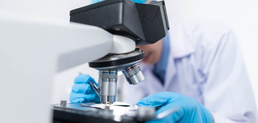
The Classical Workflow: A Day in My Previous Life
Let me paint you a picture of how things used to work. My typical day began at 7 AM, manually organizing glass slides that had accumulated overnight. The process was predictable but inefficient:
Morning Routine (2 hours):
● Sort through 40–60 glass slides from the previous day
● Check for damaged or missing slides (happened 2–3 times weekly)
● Locate corresponding requisition forms
● Prioritize urgent cases buried in the pile
● Set up multiple microscope stations
Diagnostic Phase (4–5 hours):
● Move between different microscopes for various magnifications
● Manually adjust focus and lighting for each slide
● Take notes on paper forms
● Search for comparison slides stored in filing cabinets
● Call colleagues for difficult cases (if they were available)
Reporting Phase (2–3 hours):
● Transcribe handwritten notes into digital reports
● Double-check slide numbers against reports
● Generate physical reports for delivery
● File slides in storage systems
End-of-day activities (1 hour):
● Organize slides for storage
● Update logbooks manually
● Prepare slides for courier to reference labs
This process consumed 9-11 hours daily, with frequent interruptions for slide searches, equipment adjustments, and communication delays.
The Digital Revolution: How Everything Changed
Eighteen months ago, we implemented a comprehensive digital pathology solution. The transformation has been remarkable, not just in efficiency but in the quality of our diagnostic work.
Current Workflow (6–7 hours total):
Morning Preparation (30 minutes):● Review digital case queue on my workstation
● Prioritize cases using automated urgency flags
● Check overnight AI pre-screening results
● Plan day based on case complexity and deadlines
Diagnostic Phase (4–5 hours):
● Access high-resolution digital slides instantly
● Use advanced measurement and annotation tools
● Compare current cases with archived similar cases in seconds
● Consult specialists via screen sharing in real-time
● Apply AI-assisted quantification for immunohistochemistry markers
Reporting and Review (1–2 hours):
● Generate reports directly within the digital platform
● Automatic integration with lab information system
● Digital signature and instant report distribution
● Quality assurance reviews with senior colleagues remotely
The time savings are obvious, but the qualitative improvements have been even more significant.
Quality Improvements: The Numbers Don’t Lie
Our lab tracks several quality metrics, and the improvements have been substantial:
Diagnostic Accuracy:
● Pre-digital: 94.2% concordance with reference lab opinions
● Post-digital: 97.8% concordance (measured over 1,200 cases)
● The improvement comes from better visualization tools and easier access to consultations
Turnaround Time:
● Routine biopsies: Reduced from 48–72 hours to 24–36 hours
● Urgent cases: Reduced from 4–6 hours to 90 minutes
● Cancer staging cases: Reduced from 5–7 days to 2–3 days
Error Reduction:
● Slide mix-ups: Eliminated (previously 2–3 cases monthly)
● Transcription errors: Reduced by 89%
● Lost slides: Eliminated entirely
Consultation Access:
● Specialist opinions: Increased from 12% to 67% of complex cases
● Response time: Reduced from 2–3 days to 2–4 hours
The ROI Story: Numbers That Made Management Happy
When we initially proposed digital pathology, our hospital administration was skeptical about the investment. Here's how the ROI played out over 18 months:
Direct Cost Savings:
● Slide storage reduction (repurposed space for patient care)
● Courier services (eliminated weekly slide shipments)
● Personnel efficiency: Equivalent to 1.5 additional pathologist hours daily
● Reduced repeat procedures (better initial diagnoses)
Revenue Increases:
● Increased case volume: 34% more cases with same staffing
● Premium consultation services
● Faster turnaround attracted new referring physicians
Hidden Benefits:
● Reduced pathologist fatigue and improved job satisfaction
● Better recruitment (modern facilities attract talent)
● Enhanced reputation leading to more referrals
Total ROI:187% within the first year
The subscription model made this possible without the massive upfront investment we initially feared.
The Investment Reality: Affordable Innovation
This is where I need to address the elephant in the room. When we first explored digital pathology five years ago, the quotes were staggering — ₹50–80 lakhs for basic setups, with additional costs for software, maintenance, and training.
The game-changer was finding solutions that offered subscription-based models. Instead of ₹1 Cr upfront, we started with affordable plans, which included:
● Hardware (slide scanner)
● Software platform access
● Cloud storage
● Technical support
● Regular updates
This approach allowed us to:
● Start small and scale gradually
● Include costs in operational budgets rather than capital expenditure
● Upgrade equipment as technology improved
● Receive immediate support without additional service contracts
Workflow Transformation: Before vs. After
Classical Workflow Problems We Solved:
The Lost Slide Crisis: Previously, 2–3 slides went missing weekly. Each required tissue re-cutting, re-staining, and delayed reporting. Digital slides can't get lost.
The Consultation Bottleneck: Getting specialist opinions meant packaging slides, courier services, and 3–4 day delays. Now we get consultations within hours.
The Quality Control Challenge: Manual review processes were inconsistent. Digital platforms enable standardized QC protocols with automated flagging.
The Training Dilemma: Teaching residents required gathering around single microscopes. Digital platforms allow simultaneous viewing and annotation by multiple people.
The Archive Access Problem: Finding comparison cases meant searching through years of stored slides. Digital archives provide instant access to similar cases.
Specific Workflow Innovations
AI-Assisted Pre-screening: Our system now flags potentially malignant cases for priority review. This means urgent cases get immediate attention while routine cases are batched efficiently.
Automated Measurements: For Ki-67 proliferation index, ER/PR receptor quantification, and other biomarkers, AI provides initial measurements that I verify and adjust. This reduces subjective variation and improves reproducibility.
Smart Case Routing: The system automatically routes cases to appropriate specialists based on tissue type and clinical information. Breast biopsies go to our breast pathology expert, while GI cases route to the appropriate specialist.
Quality Assurance Integration: Built-in QC protocols ensure every case meets quality standards before reporting. The system flags technical issues like poor staining or sectioning problems.
Real-World Examples from Our Lab
Case Study 1: The Emergency Consultation
Last month, a surgeon needed immediate guidance on tumor margins during a complex liver resection. Using digital pathology, I provided real-time consultation from the pathology lab while viewing the frozen section. The surgeon completed the procedure successfully without delays.
Case Study 2: The Teaching Moment
During a particularly challenging lymphoma case, I was able to simultaneously review the case with our hematology consultant in Mumbai and teach two residents. All four of us viewed the same high-resolution images while discussing differential diagnoses. This level of collaborative learning was impossible with traditional microscopy.
Case Study 3: The Quality Improvement
We identified inconsistencies in our HER2 scoring between different pathologists. Using digital quantification tools and standardized protocols, we reduced inter-observer variation from 15% to 3%, significantly improving our diagnostic reliability.
Challenges We Overcame
Initial Resistance: Some senior pathologists were skeptical about digital diagnosis accuracy. We addressed this through gradual implementation and side-by-side validation studies that demonstrated equivalent accuracy.
Technical Learning Curve: The first month involved daily technical challenges—color calibration, focus issues, and workflow adjustments. Having dedicated technical support made the difference.
Integration Complexity: Connecting with our existing lab information system took longer than expected. The key was choosing a vendor that understood healthcare IT infrastructure.
Staff Perspective: What Changed for Everyone
For Pathologists:
● Less physical strain from microscope work
● Better work-life balance (remote access for urgent cases)
● Enhanced diagnostic confidence through consultation access
● More engaging work with advanced tools
For Technologists:
● Reduced slide handling and storage requirements
● Digital tracking eliminated manual logbooks
● Quality feedback improved their staining techniques
● More consistent workflow patterns
For Residents:
● Better learning opportunities through collaborative viewing
● Access to extensive digital archives for comparison
● Exposure to advanced diagnostic tools
● More efficient knowledge transfer
The Future Roadmap
Based on our 18-month experience, here's where we're heading:
Phase 2 Expansion:
● Adding cytology digital workflow
● Implementing whole slide imaging for all cases
● Integrating molecular pathology data
Phase 3 Innovation:
● Predictive analytics for case prioritization
● Advanced AI for rare disease identification
● Telepathology services for remote locations
Practical Advice for Labs Considering Digital Transformation
Start with a Pilot: Choose one specific area (like breast pathology or GI biopsies) and demonstrate success before expanding.
Choose the Right Partner: Work with vendors who understand pathology workflows, not just imaging technology. Support quality matters more than features.
Plan for Integration: Ensure compatibility with existing systems from day one. Poor integration kills efficiency gains.
Invest in Training: Budget time and resources for staff education. The technology is only as good as the people using it.
Set Realistic Expectations: Allow 3–4 months for workflow optimization. Initial efficiency might decrease before improving dramatically.
The Bottom Line
Digital pathology has transformed our lab from a traditional, paper-based operation into a modern, efficient diagnostic facility. The workflow improvements alone justified the investment, but the quality enhancements and new capabilities have made us a regional leader in diagnostic services.
For pathologists still on the fence, my advice is simple: the technology is mature, the benefits are proven, and the investment barrier has largely disappeared. The question isn't whether to implement digital pathology, but how quickly you can do it responsibly.
The future of histopathology is digital, and from my experience leading this transformation, that future delivers better patient care through more efficient, accurate, and collaborative diagnostic workflows.
Ready to transform your histopathology lab workflow? DigiDxDoc's subscription-based digital pathology platform offers the complete solution with minimal upfront investment. Contact us to schedule a consultation and see how digital transformation can improve your lab's efficiency and diagnostic quality.